Human Back Bones Anatomy - Human Spine Anatomy High Res Stock Photo Getty Images : This framework consists of many individual bones and cartilages.. Labeled images using 3d reconstructions and an angiographic view. Most people have twelve pairs of ribs that look the same on the right and left side. A regional study of human structure. The bones, joints and walls of the pelvis. The human bones are categorized into long and short bones.
Flexible connective tissue composed of collagen and elastin fibres. Human structure and functions in health. Like other parts of our body bones keep changing all the time. There are multiple ligaments that articulate with the bones of the back and work to prevent excessive movements and strengthen the joints. The bones of the back, together, make up the vertebral column.
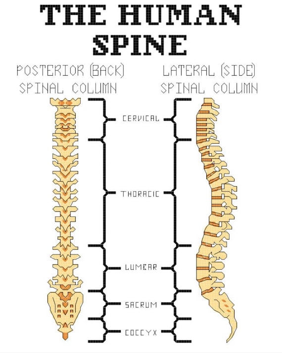
Human Spine Anatomy Cross Stitch Pattern Etsy from i.etsystatic.com Front view of muscles, back view of muscles, organs, nervous system. It's only about 3 millimeters long in an adult. There are multiple ligaments that articulate with the bones of the back and work to prevent excessive movements and strengthen the joints. .bones skeleton, human back muscles and bones, human backbone structure, pictures of human back bones, bone, human back bones diagram photos of the bone anatomy of the shoulder bone anatomy of shoulder joint, bone anatomy of the shoulder, bone structure of the human neck. An overview of the anatomy of the hand, including the bones of the hand, muscles, blood supply and nerve supply. Bone comprises the structure of the skeletal system and provides lever arms for locomotion. Flat bones are somewhat flattened, and can. The study of bone cartilage:
The feet are flexible structures of bones, joints, muscles, and soft tissues that let us stand upright and perform activities like walking, running, and jumping.
A regional study of human structure. There also are bands of fibrous connective tissue—the ligaments and the tendons—in intimate relationship with the parts of the skeleton. Anatomical diagram showing a front view of a human skeleton. Usually, the long bones have bone marrow while the short ones don't. Set of low poly models of skulls with cervical spine isolated. A collection of anatomy notes covering the key anatomy concepts that medical students need to learn. To practice basic questions and answers on all areas of human anatomy, here is complete set of 1000+ multiple choice questions scroll back to top. The vertebral column runs the length of the back and creates a central area of recession. The human skeleton provides the surface for the attachment of muscles, tendons, ligaments, etc. In this section, learn more about the anatomy of the bones of back (vertebral column). Front view of muscles, back view of muscles, organs, nervous system. In the centre of your chest. This lesson explains in detail human skeleton system.
It's only about 3 millimeters long in an adult. Human structure and functions in health. Anatomic human bones premium vector. Anatomical diagram showing a front view of a human skeleton. The bones of the back, together, make up the vertebral column.
Overview Of Skeleton Learn Skeleton Anatomy from www.visiblebody.com Cervical, thoracic and lumbar polygonal vector illustration of human skulls, front, side, and back views. The study of bone cartilage: Parts of the human skeleton. Covers the joint surfaces of mature bone. Over 3000+ pages with full illustrations and diagrams. Radiography of the hip joint : Anatomical atlas of the arteries and bones of the lower extremity: Human spine bones anatomy, sketch of skeleton backbone or vertebral column.
3d video tutorials and interactive modules on the anatomy of the vertebral column and individual vertebrae, including morphology at different levels.
In this section, learn more about the anatomy of the bones of back (vertebral column). There are multiple ligaments that articulate with the bones of the back and work to prevent excessive movements and strengthen the joints. Ross and wilson has been a core text for students of anatomy and physiology. Human neck anatomy lumbar spine disc anatomy axial skeleton anatomy bones parts of spine anatomy human backbone anatomy skeletal system back human vertebrae anatomy female back bone anatomy cervical spine anatomy diagram spine and pelvis anatomy human spinal. The human bones are categorized into long and short bones. Click on the labels below to find out more about your skeleton. The feet are flexible structures of bones, joints, muscles, and soft tissues that let us stand upright and perform activities like walking, running, and jumping. It's only about 3 millimeters long in an adult. Over 3000+ pages with full illustrations and diagrams. Set of low poly models of skulls with cervical spine isolated. Most people have twelve pairs of ribs that look the same on the right and left side. To practice basic questions and answers on all areas of human anatomy, here is complete set of 1000+ multiple choice questions scroll back to top. Parts of the human skeleton.
Parts of the human skeleton. Femur, coxal bone, acetabulum, femoral head, iliac crest. There are multiple ligaments that articulate with the bones of the back and work to prevent excessive movements and strengthen the joints. The human skeleton has a number of functions, such as protection and supporting weight. The study of bone cartilage:
Divisions Of The Skeletal System Anatomy And Physiology I from s3-us-west-2.amazonaws.com The vertebral column runs the length of the back and creates a central area of recession. The human skeleton has a number of functions, such as protection and supporting weight. 3d video tutorials and interactive modules on the anatomy of the vertebral column and individual vertebrae, including morphology at different levels. As a nurse, you will need to know the basic about the human skeleton. .bones skeleton, human back muscles and bones, human backbone structure, pictures of human back bones, bone, human back bones diagram photos of the bone anatomy of the shoulder bone anatomy of shoulder joint, bone anatomy of the shoulder, bone structure of the human neck. A regional study of human structure. Anatomical diagram showing a front view of a human skeleton. Anatomical atlas of the arteries and bones of the lower extremity:
The skull encases and protects the brain as well as the special sense organs of vision, hearing, balance, taste and smell.
Labeled images using 3d reconstructions and an angiographic view. They are attached to the spine in the back. As a nurse, you will need to know the basic about the human skeleton. Human skeleton, the internal skeleton that serves as a framework for the body. This is where layers of muscles that move the neck and back attach. Human spine bones anatomy, sketch of skeleton backbone or vertebral column. This quiz on human bones is designed to test your knowledge on the location of each individual bone. There are multiple ligaments that articulate with the bones of the back and work to prevent excessive movements and strengthen the joints. The skull encases and protects the brain as well as the special sense organs of vision, hearing, balance, taste and smell. Ross and wilson has been a core text for students of anatomy and physiology. In this section, learn more about the anatomy of the bones of back (vertebral column). Radiography of the hip joint : The bones of the back, together, make up the vertebral column.
Have you ever seen fossil remains of dinosaur and ancient human bones in textbooks, television, or in person at a museum? back bones anatomy. The bones of the back, together, make up the vertebral column.


0 Komentar